What You Should Know About Retinal Imaging
Retinal imaging helps eye specialists to detect previously undetectable indicators of eye illness. The test is harmless, and the results are simple for clinicians to understand.

By
Medical Care Review | Thursday, December 19, 2024
Stay on top of your health and well-being with exclusive feature stories on the top medical clinics and treatment centers, expert insights and the latest news delivered straight to your inbox. Subscribe today.
Fremont, CA: Retinal imaging is photographing the back of your eye digitally. It depicts the retina (where light and pictures are absorbed), the optic disc (a region on the retina that houses the optic nerve, which transmits information to the brain), and blood vessels. This assists your optometrist or ophthalmologist in detecting specific disorders and assessing the health of your vision.
Doctors have traditionally employed an instrument known as an ophthalmoscope to examine the back of the eye. Retinal imaging provides clinicians with a considerably more comprehensive digital retina image. It does not replace a conventional eye exam or dilation but adds more accuracy.
Your doctor may use special drops to dilate your eyes, broadening your scope. It will take roughly 20 minutes for your eyes to prepare for the exam.
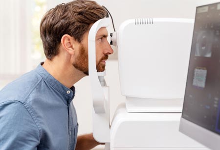
Next, lay your chin and forehead on a support to keep your head stable. While a laser scans your eyes, you will open them as wide as possible and focus directly on an item. The pictures are saved to a computer so your doctor can review them.
If the doctor suspects you have wet macular degeneration, you will likely be given a fluorescein angiography. An IV needle will be pushed into a vein in your arm to assess a dye for this test. The dye illuminates the blood vessels in your eye, allowing photographs to be taken.
The usual test lasts 5 minutes. Fluorescein angiograms take roughly 30 minutes.
If your eyes are dilated, your vision will be fuzzy for around four hours. You will also be susceptible to sunlight. You will need sunglasses and someone to drive you home.
If fluorescein dye was used, soft contact lenses should not be placed in your eyes for at least 4 hours to avoid staining them.
The test photos should be ready immediately; your doctor will usually discuss them with you before you leave.
Retinal imaging helps eye specialists to detect previously unknown indicators of eye disorders. The test is harmless, and clinicians can easily understand the data. Your doctor can keep the photos on a computer and compare them to previous scans.
Retinal imaging has limitations. It cannot identify an illness if the retina is bleeding, and it may also fail to detect issues on the retina's outer border.
Retinal imaging may be covered by medical insurance (but not vision insurance) or Medicare. It depends on the conditions of your insurance and why you're getting the test done.


